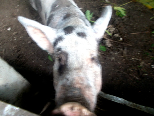
- 24 August 2019




No.Three is still Unwell


So A Technician Gives Her An Injection
of Anti-biotic

We Hope No.Three is Improving


No.Three is Lethargic and Not Eating of
Drinking Much


August 24:
One Minute Alive and the Next Minute Dead

The Work Begins

A Discovery

Work Continues


Moving the Body


The Burial


The Event:
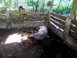
At 6.30 am feeding time No.Three had problems getting up, and had poor control over her back legs, and wouldn't eat or drink -even if we tried to give her water from a bowl. She was also panting heavily.
Around 8 am she coughed loudly and Fatima saw mucus from her mouth. Shortly afterwards she was silent. When we looked she was dead. Probably heart failure. maybe as a result of pneumonia.
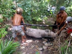
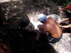
Luckily Fatima had made arrangements for a burial party -and a bunch of lads showed up when called. While digging her grave they came across bits of Butlig -which we reburied with No.Three. By 11 am it was all over.
She was a big pig -with four men struggling to move her.
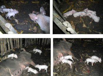
She gave us 4 live litters and a mummified one (from PPV).
We shall miss her -but life goes on.
We are suspecting infection from the chickens that roost around her pen. No.One had also been in the same pen before No.Threee moved in. We shall now have to remove some chickens because we have far too many -especially if they spread disease. But of course, we may e wrong -without skilled veterinary help it is difficult to know for certain.
We can only keep our fingers crossed that the other pigs stay healthy. We shall keep them out of the area of No.Three's pen for a while. At least until we have cleaned up and disinfected.
No.One and No.Three did not both have the same symptoms -so the cause is not simple.
---------------------
The Cause?
We think that No.One was infected by chicken poop -and that she infected No.Three.
Hopefully, we can contain the infection within the pen in which they both were housed (although not at the same time). Also the pens under the house where No.One was moved to later -allowing No.Three to be moved in to the large pen.
No.One was slaughtered, because we needed the meat -but No.Three died before we could get her treated by the animal technician.
It is possible that the problem was partly the result of the retailer of our fridge trying to fiddle us by not honoring the guarantee.
If the fridge hadn't broken down, or it had been repaired promptly -then No.One would have been slaughtered before she got ill. We suspect she had a weak immune system because she was "rescued" -after being crushed by her mother on her second day -and hand raised by us, so she did not get full immunity via her mother's milk.
Cross-infection (between species) is not so easy for a mature pig with a well developed immune system. So, it was probably necessary for No.One to be infected first (by a sick chicken?) -which perhaps made it easier for her to pass it on to No.Three.....
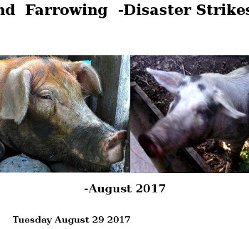
No.Three's second litter were all born dead and mummified: Suspected Porcine Parbo Virus.
Since then she did have recurring infections around her vulva. However, these seemed treatable and did not appear to affect her ability to farrow. She did have a tendency to drink lots of water.
But this is all speculation -the level of veterinary support here is not high enough to provide us with solid answers. Fatima has to do her own research.
---------------------
---------------------
The Prevention?
- Remove all Nesting Boxes from Pig Pens
- Hang up Nets to prevent chickens roosting around the Pig Pens
- Provide more roosting spaces away from Pig Pens
- Reduce Chicken Population (collect eggs, eat more chicken)
- Clean and disinfect pens before moving in new animals
- Leave pens fallow (for as long as possible) between periods of use by different animals
---------------------

Fatima's Final Researched Diagnosis:
No.Three (a 4-year old multiparous sow)
July 30, only 27 days before she died, No.Three was separated from her piglets and moved to the North Garden Pen. As expected, 4 days after weaning, she started coming in heat. She showed no signs of illness.
August 16, she started vocalizing as if in heat. This was unusual because she was not supposed to be in heat again. She seemed to eat and drink normally. However, I detected some other subtle changes in her behaviour, for example, lying down too soon after eating and not finishing all her water like she usually did. 5 days later, I noticed that No.Three didn't finish her food from yesterday and she had elevated temperature. There was also some blood and clear liquid in her vulva which she has had on several occasions. However, because she had a temperature and was off her food and water, her condition was something to worry about. She would eat several bananas and drink coconut water but not very much. Her skin, particularly the insides of her legs and chest, had a pinkish tinge. There were no obvious signs of cyanosis or purple or blue colour of the ears and extremities.
August 22, we called the animal technician to inject Penicillin-Streptomycin in the morning. 8 hours later, she remained lethargic, inappetant and some gurgling sounds can be heard when she breathed. Two days later, At 5:30 in the morning, I could hear very loud deep coughing. She had difficulty breathing, mucus was dripping from her nose and mouth. She moved frequently although very weak, trying to find a way to breathe. At the last moment, gasping for air, she trembled from head to tail and fell down dead.
No.Three died at around 8:30 in the morning.
Diagnosis
Acute Pleuropneumonia, infection by strains of the bacterium Actinobacillus pleuropneumoniae (APP)
Note: In August 29, 2017, No.Three farrowed to 2 stillborn piglets and 6 mummified fetuses. Our suspicion was Porcine Parvovirus (PPV). The prolonged gestation period of 117 days was a sign that something wasn’t right. Note also that a few weeks into gestation, No.Three had a slightly bloody discharge for several days. This could’ve been an early sign of infection.
Pneumonic pigs are often fevered and appear reddened. They become inappetant and fail to drink. They are weak and respiratory rates are raised. Harsh lung sounds, bubbling sounds and tracheal sounds can all be heard. Damage to the lungs leads to increased load on the heart. In the peracute and acute form, before they die, pigs may have severe breathing difficulties, coughing, with open-mouth breathing and a foamy, blood-tinged discharge from the nose and mouth.
Pleuropneumonia is caused by Actinobacillus pleuropneumoniae (APP), a Gram negative rod-shaped bacterium which produces fimbriae (filaments), a protective capsule and at least four toxins (ApxI, ApxII, which are haemolytic and destroy red blood cells, ApxIII which kills cells and Apx IV, produced in vivo and essential for pathogenicity).
At least 15 different capsular types (serotypes) occur, but each serotype may not produce all the toxins. Different combinations of serotypes are found in each country. The organism colonises the tonsils and adheres to them, but in non-immune pigs it is inhaled into the lungs where it adheres to cells lining the alveoli. There it is taken up by alveolar macrophages (lung defence cells). Lung defence cells cannot kill it because of the capsule. Thus, the cells are killed WITHIN AN HOUR by the toxins. The organism multiplies in the undefended lung and produces more toxin which damages the lung, causing inflammation (pneumonia, leakage of capillaries (leading to pleurisy) and destruction of the lung tissue, some of which damage is irreversible. If the pig recovers, it develops protective immunity to infection, particularly to infecting serotype. Infection may be inapparent, but 15-30% of pigs may suffer depression, inappetence, high fever (41.5°C, 107°F), or laboured breathing, especially after rising to drink of after disturbance within hours of infection. Cyanosis (a purple tinge of the ears and feet), subnormal rectal temperature and death may follow within 4-6 hours of the onset of clinical signs.
Although primarily a disease of growing pigs, APP infection may be fatal in adults or cause sows to abort. The course of the disease varies from peracute to chronic. The presence of pigs with respiratory distress, coughing and exercise intolerance in very poor condition with some mortality may suggest chronic disease. The diagnosis can be confirmed in most cases by post-mortem examination.
Once established in a herd, the disease may be evident only as a cause of reduced growth rate and pleurisy at the abattoir, although acute disease exacerbations may occur. Concurrent infection with mycoplasma, pasteurellae, porcine reproductive and respiratory syndrome, or swine influenza virus is common. It must be stressed that respiratory disease problems in pigs are frequently the result of multiple agents (co-infection) and rarely due to the effects of a single pathogen.
Treatment and Control
Rapidity of onset and persistence in infected herds make treatment difficult. Ceftiofur, tilmicosin, tetracyclines, synthetic penicillins, tylosin, and sulfonamides have been used. The first treatment should be parenteral, followed by medication given in water or feed, which may also protect contact pigs.
Because survivors frequently remain carriers, control is difficult, although good results are being claimed for some vaccines. Segregated early weaning, “all-in/all-out” management, reduced stocking rates when possible, and improved ventilation are recommended. In herds free of the disease, replacements should be purchased from herds free of APP. If the disease proves difficult to control, herd depopulation and repopulation should be considered. Serologic testing effectively detects previously infected herds but may not identify carrier animals.
It is difficult to keep herds free of respiratory diseases. Aerosols have been suspected as sources of pathogen entry onto naive farms. Organisms such as Mycoplasma hyopneumoniae have been postulated to be transmitted over distances of as far as 2 miles, depending on climate, terrain, and density of pigs in the locality; however, this assumption is based on speculation and use of mathematical models rather than on experimental data.
Closed herds may help to establish immunity. This entails not buying in any live animals, using artificial insemination or embryo transfer to bring in new genetic material. This helps to establish immunity to present organisms and avoid introduction of new infections, strains, or serotypes.
Respiratory disease is endemic in many herds. The main control factors are stress management, stocking density, ventilation, temperature control, and freedom from mixing or moving. Multiple site production or an “all-in/all-out” policy, in which the entire barn or air space is emptied before refilling, can very effectively minimize the potential effect of chronic pneumonia. This greatly decreases the need for preventive and therapeutic medication.
Actinobacillus pleuropneumoniae does not affect humans and has no public health significance.
Notes on Route of Infection
1. No.Three may have acquired the infection while she was in the North Garden Pen where No.One has recently vacated. The pen wasn't thoroughly cleaned, dried and disinfected. No.Three was moved to the pen just barely 2 days after No.One vacated it.
"Pleuropneumonia infection is by the respiratory route from contact between pigs, by aerosol, from contaminated drinkers and from contaminated clothing etc. The organism does not survive for long on drying, but can persist in clean water for days. Introduction to a clean farm is usually by means of carrier pigs."
2. No.One may have acquired the infection as a piglet. No.One was rescued from crushing at 2 days of age. She was fed sow's milk substitute. She joined her siblings when they were weaned. This was probably when she became infected. At slaughter, No.One's lungs showed signs of pleuropneumonia. No.One had no obvious clinical signs of pneumonia (no obvious respiratory distress). However, she was inappetant at some times, particularly when changing feed, which may have led to gastric ulcer. This was evident at slaughter: lung lesions and some pleurisy.
"It has been suggested that only a few piglets get infected from their mother during the lactation period when the level of maternal immunity is usually high, and that these infected pigs spread the bacteria to other pigs after weaning, when passive immunity declines. The persistence of protective colostral antibodies in piglets may range from 2 to 10 weeks postpartum, depending on the initial level. Early weaning has been used with a relatively high level of success to produce APP-free piglets from infected sows."
3. No.One may have had chronic plueropneumonia and No.Three got infected with acute pleuropneumonia. No.Three's history of Porcine Parvovirus two years back may or may not be a contributing factor. Stress (the wean-to-oestrus interval is one of the most stressful times in a sow's life) and fluctuating weather conditions may be aggravating circumstances.The prevalence of infection may also vary greatly depending on the serotype of the bacterium.
"Some serotypes are highly contagious but poorly virulent (e.g., serotype 12, 6, 8 and 15) whereas the contrary is true for others (e.g., serotypes 1 and 5). Morbidity and mortality depend on the virulence of the strain and on the environment. Factors such as overcrowding and adverse environmental conditions may promote the development and spread of the disease. Co-infections with other respiratory pathogens may also increase clinical signs and mortality."
Can chickens and ducks be a source of infection?
Animal species other than pigs such as birds, rodents or insects have NOT been shown to serve as reservoirs or significant vectors of APP. Survival in the environment is considered to be of short duration. However, when protected with mucus or other organic matter, APP can survive for several days or weeks. This may explain why fomites and people are occasionally involved in transmission.
However, an APP isolate with characteristics of some Pasteurellaceae family members (Gram-negative bacterium, pleomorphic, and NAD-dependent) was isolated from layer hens showing clinical signs of infectious coryza ( a serious bacterial disease of chickens which affects respiratory system and it is manifested by inflammation of the area below the eye, nasal discharge and sneezing). This bacterium indicated 99% identity with A. pleuropneumoniae serotypes 3 and 7. (From "Isolation of Actinobacillus pleuropneumoniae from Layer Hens Showing Clinical Signs of Infectious Coryza" by V. M. Pérez Márquez, et al, June 2014).
The three porcine diseases that are definitely transmitted by birds are avian tuberculosis, transmissible gastro-enteritis (TGE) and erysipelas, although it is likely that other infectious agents such as PRRS virus may be carried on birds' feet or pass through their alimentary tracts into their droppings.
Eradication
Owners of infected herds may attempt eradication of the organism. Several procedures have been successful but a careful economic evaluation is recommended before starting an eradication program. Depopulation and restocking with pigs from certified App-free herds is one effective means by which to eliminate the organism. This method is usually implemented in commercial herds which are facing severe Porcine Pleuropneumonia problems as well as other health issues (e.g., PRRS). Nucleus herds have to use other methods to preserve their blood lines. One approach that has succeeded in the past is medicated early weaning, as APP was shown to be a relatively late colonizer in piglets. Certain serotypes of APP have been reported as successfully eliminated using serological testing, antibiotic treatment and partial depopulation (e.g., 1, 5, 7). However, it should be emphasized that antibiotic treatment can not completely eliminate APP from carrier animals. Thus antibiotic treatment is only a tool to reduce transmission of APP during the eradication process.
References:
Porcine pleuropneumonia (PPP) caused by Actinobacillus pleuropneumoniae (App)
<http://porkgateway.org/resource/porcine-pleuropneumonia-ppp-caused-by-actinobacillus-pleuropneumoniae-app/>
Pleuropneumonia (app)
<https://www.pigprogress.net/Health/Health-Tool/diseases/Pleuropneumonia-app/>
Pleuropneumonia in Pigs
<https://www.msdvetmanual.com/respiratory-system/respiratory-diseases-of-pigs/pleuropneumonia-in-pigs>
Overview of Respiratory Diseases of Pigs
<https://www.merckvetmanual.com/respiratory-system/respiratory-diseases-of-pigs/overview-of-respiratory-diseases-of-pigs>
Pneumonia
<https://www.pigprogress.net/Health/Health-Tool/diseases/Pneumonia/>
Isolation of Actinobacillus pleuropneumoniae from Layer Hens Showing Clinical Signs of Infectious Coryza
<https://www.jstor.org/stable/26431761>
Airborne transmission and other methods
<https://thepigsite.com/disease-and-welfare/managing-disease/airborne-transmission-and-other-methods>
---------------------
---------------------
Piglets 2015
Butlig's
Operation
Butlig,s
Diary
---------------------
